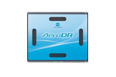

WAYNE, N.J., Nov. 11, 2015 /PRNewswire/ -- Konica Minolta, a market leader in medical diagnostic Primary Imaging Solutions, will feature AeroDR LT,* its new digital radiology solution, at the 2015 Annual Meeting of the Radiological Society of North America (RSNA) Booth #2729. The AeroDR LT provides radiologists with the state-of-the-art capabilities of the AeroDR XE in a lower weight version to deliver Primary Imaging at the point-of-care.

The AeroDR LT was designed to maximize efficiencies by minimizing the effects of wear and tear that come with alternative wireless flat panels. The lightweight, hardened, IPX6-rated, liquid resistant enclosure allows radiologists to help optimize patient outcomes, productivity and return on investment both in-room and outside of the radiology department, such as at the patient's bedside or in ER/trauma or intensive care/critical care units. The AeroDR LT weighs 5.5 pounds (2.5 kg) and has the ability to hold patients up to 661 pounds (300 kg) on a bed.
Advanced digital features, such as compatibility with AeroSync automatic exposure detection and roaming capabilities, and usability with most fixed and portable X-ray devices, are the cornerstone of the modern radiology department. Whether on the go or in the treatment room, radiologists can diagnose a patient with AeroDR LT's ability to preview images within one second of use and deliver fully processed images in as little as six seconds.
Customers can leverage the AeroDR LT to help streamline workflow and avoid unplanned downtime. The lithium ion capacitor lets users receive up to 4.1 hours of imaging use or 150 images with only a 13-minute charge time. The reliable built-in panel drop sensors and continuous panel handling data monitoring can help reduce repair costs and avoid catastrophic failures.
"Konica Minolta's next generation of digital radiography flat panel solutions, the AeroDR LT, is an ideal solution for radiologists, particularly those practicing in smaller facilities, who are considering upgrades to the medical equipment," said Guillermo Sander, Senior Product Manager, Digital Radiography, Konica Minolta Medical Imaging. "With this one investment, radiologists can digitally transform their imaging capabilities and data transfer needs, while making value-based decisions."
About Konica Minolta Medical Imaging
Konica Minolta Medical Imaging is a world class provider and market leader in medical diagnostic Primary Imaging. With over 75 years of endless innovation, Konica Minolta is globally recognized as a leader providing cutting-edge technologies and comprehensive support aimed at providing real solutions to meet customer's needs. Konica Minolta Medical Imaging, headquartered in Wayne, NJ, is a unit of Konica Minolta, Inc. (TSE: 4902). For more information on Konica Minolta Primary Imaging Solutions, please visit www.konicaminolta.com/medicalusa and follow us on Twitter.com/KonicaMinoltaMI.
|
Company name |
KONICA MINOLTA, INC. |
|
Headquarters |
JP TOWER, 2-7-2 Marunouchi, Chiyoda-ku, Tokyo, Japan |
|
Founded
|
December 1936 |
|
FY 2014 Revenue |
$8.5 Billion |
|
Number of Employees |
Approx. 41,600 (2015) |
|
Business Lines
|
The Konica Minolta Group operates in sectors ranging from business technologies, where our products are typified by MFPs (multi-functional peripherals), and Industrial Business (former Optics Business), where our products include pickup lenses for optical disks, and TAC film, a key material used in LCD panels, to healthcare, where we make digital X-ray diagnostic imaging systems. |
*Static loads have no effect on images and detector even when applied directly. The measurement method based on Konica Minolta's standards. The maximum patient weight performance of this product does not guarantee that product damage or failure will not occur. The product may fail to maintain its waterproof performance (equivalent to IPX6) if it has been dropped.
The waterproof performance of this product does not guarantee that product damage or failure will not occur.
The performance may vary depending on component configurations and usage environments. The performance described here is obtained during exposures with an X-ray generator. Three exposures per examination; in a five-minute cycle examination (with the positioning time assumed of 20 seconds); exposures with an X-ray generator.
The performance described here is expected when fully charged. The performance level may fluctuate depending on the usage environment and frequency of use (the ability to always obtain the performance described here is not guaranteed).
